

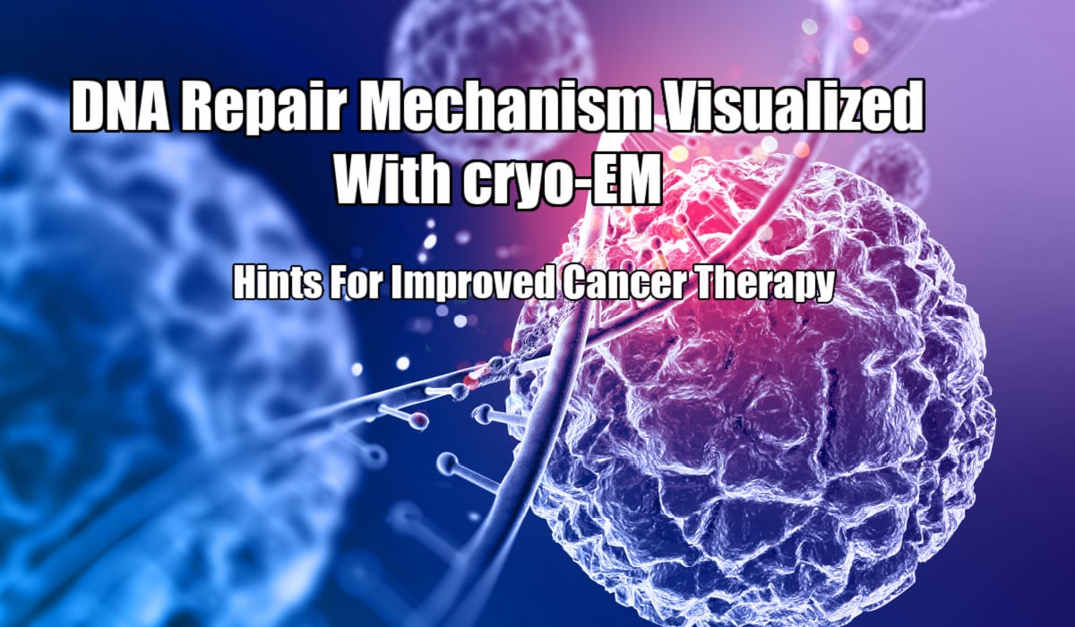
cryo-EM Visualization Of DNA Repair Mechanism

From cancer therapy to toxins, radiation, and sunlight can seriously damage DNA in both healthy and harmful cells. Even though the body has advanced to efficiently restore and treat damaged cells, the dynamics that enable this natural repair remain unknown.
In a new study, scientists from Northwestern University have employed cryo-EM (Cryogenic Electron Microscopy) to observe this process by irradiating the peculiar cycle of DNA damage sensing and repair. The scientists assume this new detail could possibly form the foundation for discerning how cells react to radiation and chemotherapy and possibly even result in improved cancer treatments.
The work was published in the journal Nature on April 14, 2021. The study provides a new understanding of how proteins function together to detect and repair DNA DSB (Double-Strand Breaks).
When a DSB-the most severe version of DNA damage-happens, the repair pathway might delete or insert genes at the site of breakage or possibly rearrange genes across the strand. In cases of chromosomal rearrangements, destructive changes can result in the development of cancer.
Scientists can procure 3-D images of macromolecular complexes at atomic resolution using cryo-EM. Based on Yuan He, assistant, Molecular Bioscience, Weinberg College of Arts & Sciences, the ability of cryo-EM to form images of dynamic machinery goes way beyond the potential of other structural biology approaches.
Imaging DNA-protein structures at different transitory phases enabled Yuan He’s group to detect and devise a prototype of the cell’s repair pathway.
In the video, when the bottom element of the DNA-protein complex comes under stress, DNA strands are held to close proximity; once the element goes to a relatively relaxed state, the movement then seals the 2 DNA strands together.
Yuan He, the corresponding writer of the study stated that there are several aspects that function together to repair the breakage. They are taking the most direct method to resolve the issue by concentrating on proteins as they detect and repair a break.
The resulting model demonstrated that, occasionally, 2 copies of DSB sensing complexes can bridge and hold together DSBs while the complexes summon other factors to the site of breakage. In another crucial state, proteins line up the 2 DNA strands for a ligase enzyme to come and repair the breakage. The lab later suggested a prototype of this pathway, demonstrating how DNA aligns and bridges as it shifts between states.
Seeing is believing is a popular saying within molecular biology, and is the one Yuan He applied to his study directly. By visualizing the elegant process directly, a viewer can easily make links and discern how the complex system functions jointly. However before this methodology was accessible, Yuan He claimed that it was alike to a blind man identifying an elephant by touch.
Siyu Chen, the first writer of the paper and postdoctoral fellow at Yuan He’s lab, asserted that discoveries are typically thought of as something complex and big. The question answered is basic and direct: How proteins join the broken DNA together?
The laboratory believes the results have indications for how our cells revert and react to chemotherapy and radiation. Upcoming studies could bring additional targeted treatments for this newly studies pathway.
The article is titled ‘Structural basis of long-range to short-range synaptic transition in NHEJ’.
Breakthrough For Improved Cancer Treatment, Proteins Recognize DNA Breaks, cryo-EM Visualization Of DNA Repair, Proteins Recognize DNA Breaks, Understanding How DNA Repairs, Breakthrough For Improved Cancer Treatment
Also Read: Researchers Discover Direct Proof Of Evolution Of Venom Glands From Salivary Glands


